ModalityCRDX
X-ray (radiography)
A projection image made with a very short X-ray exposure. A study usually has one to four large grayscale DICOM images.
On this page
An X-ray image, or radiograph, is the oldest and most common type of medical imaging. A short burst of X-rays passes through the body and creates a flat image on a detector. Bones look white, air looks black, and soft tissues appear in shades of gray.
Key facts
| Fact | Details |
|---|---|
| DICOM modality code | CR (computed radiography) or DX (digital radiography) |
| Typical number of images | 1 to 4, sometimes more |
| Typical study size | 5 to 60 MB (5 to 20 MB per uncompressed image) |
| Exam duration | 5 to 15 minutes; the exposure lasts a fraction of a second |
| Radiation | Yes, low; from less than 0.001 mSv (hand) to about 1.4 mSv (lumbar spine) |
| Contrast agent | None for plain radiographs |
How it works
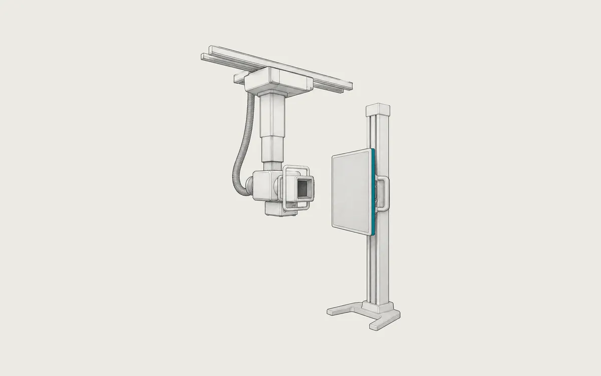
An X-ray tube sends a short pulse of X-rays through the part of the body being examined. Bone absorbs more X-rays than soft tissue, and air absorbs almost none. A detector on the other side records the X-rays that pass through.
Two digital technologies are common. In computed radiography (CR), the image is stored on a phosphor plate in a cassette, and a reader scans the plate with a laser. In digital radiography (DR), a flat-panel detector produces the image within seconds. Both have replaced film in most hospitals.
X-ray compared with CT
A radiograph is a projection: all structures along the X-ray path overlap in one flat image. For this reason, doctors often order two views at right angles, such as front and side. CT rotates the X-ray tube around the body and calculates separate slices without overlap. CT shows much more detail, especially in soft tissues, but its dose is much higher. For example, a chest CT gives about 60 times the dose of a chest X-ray.
Uses
Common uses include:
- fractures, dislocations and joint problems such as arthritis
- the chest, for example the lungs, heart size, pneumonia, and the position of tubes and lines
- the abdomen, for example when a bowel obstruction or a swallowed object is suspected
- the spine, including curvature (scoliosis)
- bone age and growth in children
Special types of X-ray imaging have their own articles: mammography, dental X-rays, angiography and fluoroscopy and bone density scans.
Preparation and what to expect
Most radiographs need no preparation. You may be asked to remove clothing, jewelry, glasses and other metal objects from the area and to wear a gown. Tell the staff if you are or might be pregnant.
The technologist positions you standing, sitting or lying. You must not move during the exposure, and for a chest X-ray you hold your breath for a moment. The technologist steps behind a screen to start the exposure, and you feel nothing. The images appear on a monitor within seconds and are checked before you leave. A radiologist then sends a report to your doctor.
Safety
Radiographs use ionizing radiation, but the doses are low. RadiologyInfo lists typical effective doses for adults of about 0.1 mSv for a chest X-ray, 1.4 mSv for the lumbar spine, and less than 0.001 mSv for a hand or foot. Natural background radiation is about 3 mSv per year, so a chest X-ray equals about 10 days of background radiation. The actual dose depends on the protocol, the equipment and the size of the patient.
The risk from such doses is very small. Exposures for children are adjusted to their size. Lead shields are used less often than in the past, because a shield can hide important parts of the image; practice differs between hospitals and countries. If you are pregnant, the doctor will decide whether the X-ray is needed now or can wait.
What an X-ray study looks like in DICOM
Images from phosphor plate systems have the modality CR and the SOP class Computed Radiography Image Storage (1.2.840.10008.5.1.4.1.1.1). Flat-panel systems use the modality DX and the SOP class Digital X-Ray Image Storage - For Presentation (1.2.840.10008.5.1.4.1.1.1.1), the processed image that doctors read. Digital X-Ray Image Storage - For Processing (1.2.840.10008.5.1.4.1.1.1.1.1) holds less processed detector data for computer analysis and is rarely archived. Some digital systems send their images as CR objects for compatibility with older archives.
Each image is usually one file. Typical sizes are about 2000x2500 to 3000x3000 pixels or more, usually with 16 bits allocated and 10 to 16 bits used.
Photometric Interpretation (0028,0004) matters for radiographs. With MONOCHROME2, low values are displayed dark. With MONOCHROME1, low values are displayed bright, and many CR and DX systems use this option. A program that ignores this attribute shows the image as a negative.
Other useful tags:
- View Position
(0018,5101), for examplePA,APorLL(left lateral) - Body Part Examined
(0018,0015), for exampleCHESTorHAND - Image Laterality
(0020,0062),RorL - KVP
(0018,0060)and Exposure(0018,1152)in mAs - Exposure Index
(0018,1411)and Deviation Index(0018,1413), which show whether the detector received the intended exposure - Imager Pixel Spacing
(0018,1164), the pixel size at the detector
Body parts are slightly magnified on the detector, so a distance measured with Imager Pixel Spacing is slightly larger than the real distance in the body.
How to view the images
Any DICOM viewer can open a radiograph. You can zoom, adjust brightness and contrast (the window), and measure distances and angles. Treat measurements as approximate unless the image was calibrated. Exporting to JPEG keeps only 256 shades of gray and can lose fine detail (see converting DICOM).
Weasis (free, open source), MicroDicom (free for non-commercial use, Windows) and Horos (free, open source, macOS) can display radiographs. See how to open DICOM files.