ModalityPT
PET and PET/CT
A radioactive tracer shows how active tissues are, and CT shows where they are. A viewer can fuse the PET (PT) and CT series into one picture.
On this page
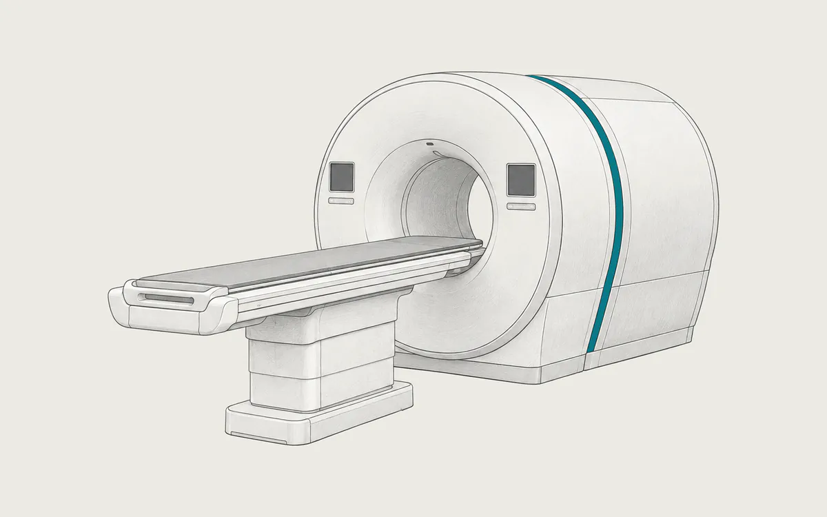
Positron emission tomography (PET) shows how active tissues are. A small amount of a radioactive tracer is injected into a vein, and the scanner measures where it collects. Most scanners combine PET and CT in one machine (PET/CT), so the activity map can be placed over detailed anatomy. In DICOM, the study contains PET images (PT) and CT images (CT).
Key facts
| Fact | Details |
|---|---|
| DICOM modality code | PT for PET; CT for the CT part of the same study |
| Typical number of images | Several hundred to a few thousand |
| Typical study size | 100 MB to 1 GB, mostly CT images |
| Exam duration | About 2 hours in total, including about 30 minutes of scanning |
| Radiation | Yes, from the tracer and the CT; often about 5 to 25 mSv in total |
| Contrast agent or tracer | A radioactive tracer, most often FDG; sometimes iodine-based contrast for the CT |
How it works
The most common tracer is FDG (fluorodeoxyglucose), a modified sugar labeled with radioactive fluorine-18, which has a half-life of about 110 minutes. Cells that use a lot of sugar, such as many cancer cells, take up more FDG. Other tracers target specific diseases, for example PSMA tracers for prostate cancer.
The tracer releases positrons. Each positron soon meets an electron, and both turn into two gamma rays that fly in opposite directions. A ring of detectors records pairs that arrive at the same time, and a computer reconstructs 3D images from millions of such events. The CT scan, done just before the PET scan, shows anatomy and is used to correct the PET data for gamma rays absorbed in the body (attenuation correction).
Uses
- finding cancer, staging it, and checking the response to treatment or a return of the disease
- blood flow to the heart and whether damaged heart muscle is still alive
- brain disorders, such as memory loss, seizures and tumors
- some infections and inflammatory diseases
FDG also collects normally in the brain, heart, kidneys and bladder (the last two because it leaves the body in urine), so a bright area does not always mean disease.
Preparation and what to expect
For an FDG scan you usually must not eat for several hours, and drinks with sugar are not allowed; water is encouraged. Many centers ask you to avoid hard exercise the day before. People with diabetes receive special instructions, because blood sugar changes how FDG spreads. Tell the staff about your medicines and allergies, and arrive on time, because the tracer decays quickly.
After the injection you rest quietly for about 60 minutes, then empty your bladder and lie on the table. You lie still while the CT takes less than two minutes and the PET scan about 20 to 30 minutes. Afterwards you can usually return to normal activities; drinking water helps clear the tracer.
What SUV means
Reports often give standardized uptake values (SUV), for example "SUVmax 6.2". SUV compares the tracer concentration in an area with the value expected if the tracer were spread evenly through the body, so 1 means average uptake. No single value separates cancer from other causes, such as inflammation. SUV also depends on timing, blood sugar, body weight, the scanner and the reconstruction, so values are best compared between scans with the same protocol. Ask your doctor what the values mean for you.
Safety
RadiologyInfo lists about 22.7 mSv for a whole-body PET/CT protocol, equal to about 7.6 years of natural background radiation (about 3 mSv per year). The total is lower when the CT is a low-dose scan and higher when a full diagnostic CT is added. The dose depends on the protocol, the machine, the injected tracer activity and body size.
Allergic reactions to PET tracers are extremely rare, and most of the radioactivity is gone within a day. During pregnancy, PET/CT is done only when the benefit clearly outweighs the risk, so tell the staff if you are or might be pregnant. If you are breastfeeding, ask in advance; you may need to pause for a short time, depending on the tracer. For iodine-based contrast, see CT.
What a PET/CT study looks like in DICOM
PET series have Modality PT and use Positron Emission Tomography Image Storage (1.2.840.10008.5.1.4.1.1.128), usually one file per slice. Newer systems may use the multi-frame Enhanced PET Image Storage (1.2.840.10008.5.1.4.1.1.130). CT series use CT Image Storage (1.2.840.10008.5.1.4.1.1.2). A study usually holds a low-dose CT and two PET series, with and without attenuation correction. Some also include a diagnostic CT or a rotating maximum intensity projection (MIP). Fused views saved as secondary capture images are pictures only and cannot be measured. PET and CT usually share one Frame of Reference UID (0020,0052), so a viewer can fuse them.
PET slices are usually 128x128 to 256x256 pixels, CT slices 512x512. PET pixels are 16-bit integers with a separate Rescale Slope (0028,1053) for each image. Rescaled values are usually in becquerels per milliliter, marked BQML in Units (0054,1001).
To calculate SUV, a viewer needs:
- Radiopharmaceutical Information Sequence
(0054,0016)with Radionuclide Total Dose(0018,1074)in becquerels, Radiopharmaceutical Start DateTime(0018,1078)or the older Radiopharmaceutical Start Time(0018,1072), and Radionuclide Half Life(0018,1075) - Patient's Weight
(0010,1030) - Units
(0054,1001)and Decay Correction(0054,1102), usuallySTART
If a value is missing or wrong, SUV is wrong or not shown. Basic de-identification removes Patient's Weight unless patient characteristics are kept, so SUV can be lost after de-identification.
How to view the images
PET is shown in color or in inverted gray, and fused views place color PET over gray CT. Weasis (free, open source), Horos (free, open source, macOS), OHIF Viewer (free, open source, in a web browser) and RadiAnt (commercial, Windows) support fusion and SUV. The report, not the colors on screen, gives the medical interpretation. See how to open DICOM files.