ModalityMG
Mammography and breast tomosynthesis
Low-dose X-ray imaging of the breast. A standard study has four very large images; tomosynthesis adds stacks of thin slices and can reach several gigabytes.
On this page
Mammography is an X-ray examination of the breast. It is used for screening, which means looking for breast cancer in people without symptoms, and for diagnosis. Digital breast tomosynthesis (DBT), also called 3D mammography, shows the breast as a stack of thin slices.
Mammograms have a very high resolution, so their DICOM files are among the largest in medical imaging.
Key facts
| Fact | Details |
|---|---|
| DICOM modality code | MG |
| Typical number of images | 4 for a standard study (two views of each breast); tomosynthesis adds tens of slices per view |
| Typical study size | 2D: about 50 to 250 MB without compression; with tomosynthesis: several hundred MB to a few GB |
| Exam duration | About 30 minutes; each compression lasts a few seconds |
| Radiation | Yes, low; about 0.3 mSv for a screening study |
| Contrast agent | None for standard mammography |
How it works
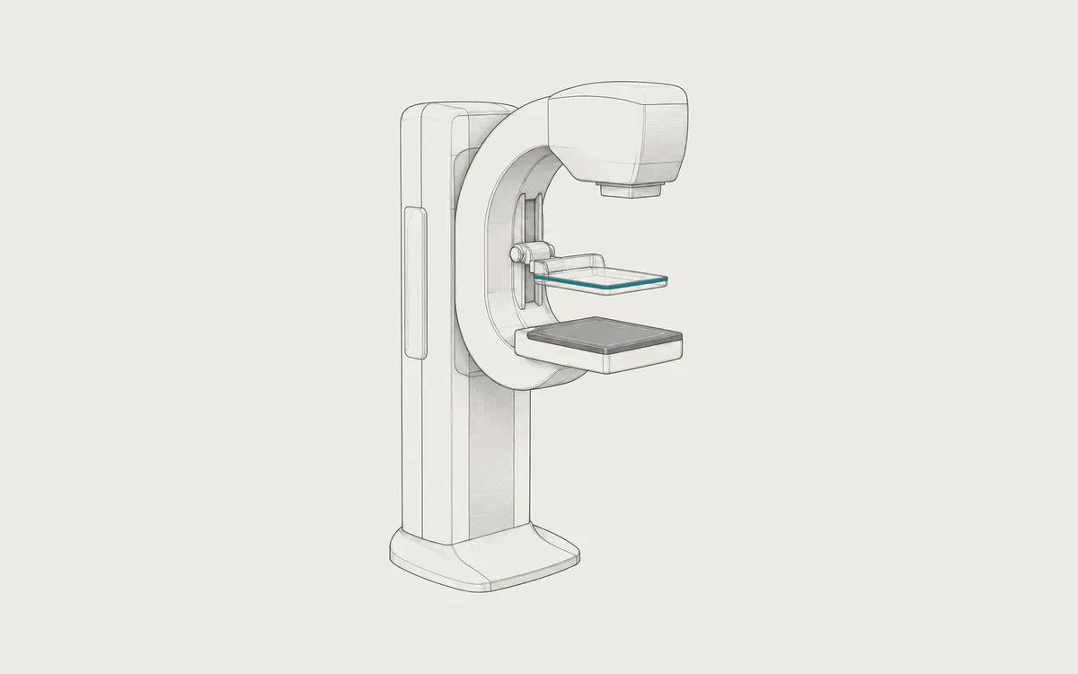
A mammography unit uses low-energy X-rays and a high-resolution detector. The breast is placed on a support and pressed by a clear plastic paddle. Compression spreads the tissue, reduces blurring from movement and allows a lower dose. The two standard views are craniocaudal (CC), taken from above, and mediolateral oblique (MLO), an angled side view.
In tomosynthesis, the X-ray tube moves in an arc over the compressed breast and takes a series of low-dose images from different angles. A computer reconstructs them into slices, usually about 1 mm apart. Looking through the slices reduces the problem of overlapping tissue, which can hide a lesion or look like one. Many systems also calculate a synthetic 2D image from the tomosynthesis data, so a separate 2D exposure is not always needed.
Uses
Common uses include:
- screening for breast cancer, at ages and intervals that differ between countries and organizations
- examining a lump, pain, nipple discharge or skin changes
- checking a finding from screening, often with extra views or ultrasound
- guiding a needle biopsy
- follow-up after breast cancer treatment
Reports often state breast density. Dense tissue can hide cancer on a mammogram, and some patients are offered additional tests, such as ultrasound or MRI.
Preparation and what to expect
On the day of the exam, do not use deodorant, powder or lotion under the arms or on the breasts. They can appear on the images as small white spots that look like calcium deposits. If your breasts are tender before your period, schedule the exam for the week after it. Tell the staff about breast implants, previous breast surgery, pregnancy or breastfeeding. If you had mammograms elsewhere, bring the previous images, ideally as DICOM files, because comparison with earlier images is very important.
You undress above the waist. The technologist positions each breast and compresses it for a few seconds while the image is taken. Compression can be uncomfortable or painful, but it lasts only a short time. Tell the technologist if the pain is strong. Tomosynthesis uses the same positions and compression.
Safety
Mammography uses a low dose of ionizing radiation. RadiologyInfo lists typical effective doses of about 0.28 mSv for screening digital mammography and 0.34 mSv for screening tomosynthesis. Natural background radiation is about 3 mSv per year, so a screening study equals about five to six weeks of background radiation. The actual dose depends on the equipment, the protocol and the size and density of the breast. Tomosynthesis combined with a separate 2D exposure gives a higher dose than either one alone.
Screening has known limits. Some findings turn out to be harmless after extra tests (false positives), some cancers are missed, and some cancers that are found would never have caused harm (overdiagnosis). Ask the referring doctor about the benefits and limits of screening for you.
What a mammography study looks like in DICOM
Mammograms have the modality MG. 2D images use Digital Mammography X-Ray Image Storage - For Presentation (1.2.840.10008.5.1.4.1.1.1.2), the processed image for reading. Digital Mammography X-Ray Image Storage - For Processing (1.2.840.10008.5.1.4.1.1.1.2.1) holds less processed data, mainly for computer-aided detection (CAD), and is not always archived.
Tomosynthesis slices use Breast Tomosynthesis Image Storage (1.2.840.10008.5.1.4.1.1.13.1.3): usually one multi-frame file per view, with each slice as a frame. The synthetic 2D image is usually stored as an ordinary 2D mammogram. The projection images, if kept, may use Breast Projection X-Ray Image Storage or vendor-specific objects. Some older systems stored tomosynthesis in vendor-specific formats that general viewers cannot display.
Why the files are so large
A 2D mammogram typically has between about 2400x3000 and 4800x6000 pixels, with a pixel size of 50 to 100 micrometers and 16 bits allocated per pixel. One uncompressed image takes tens of megabytes. A tomosynthesis view contains tens of slices of similar size, so a full study can reach several gigabytes. Lossless compression often halves this.
Useful tags:
- Image Laterality
(0020,0062),RorL - View Position
(0018,5101), which often containsCCorMLO, and View Code Sequence(0054,0220) - Body Part Thickness
(0018,11A0), the compressed breast thickness in millimeters, and Compression Force(0018,11A2)in newtons - KVP
(0018,0060)and Anode Target Material(0018,1191) - Organ Dose
(0040,0316), which in mammography is the estimated mean glandular dose in dGy
Results of CAD software can be stored as a Mammography CAD SR, and many systems also create an X-Ray Radiation Dose SR.
How to view the images
Radiologists read mammograms on high-resolution medical displays, often 5 megapixels, with the right and left breasts side by side and earlier studies for comparison. A laptop or phone cannot show every fine detail.
Most DICOM viewers, such as Weasis (free, open source), Horos (free, open source, macOS) and MicroDicom (free for non-commercial use, Windows), open 2D mammograms. If a viewer shows only the 2D images, it may not support the tomosynthesis objects. See how to open DICOM files.