ModalityCT
CT scan (computed tomography)
Cross-sectional X-ray imaging of any part of the body. One study holds hundreds to thousands of DICOM slices with values in Hounsfield units.
On this page
A CT scan (computed tomography, also called a CAT scan) uses X-rays and a computer to make cross-sectional images of the body. Each image shows a thin slice, like one slice of a loaf of bread.
Key facts
| Fact | Details |
|---|---|
| DICOM modality code | CT |
| Typical number of images | 100 to several thousand, in several series |
| Typical study size | 50 MB to 2 GB (about 0.5 MB per uncompressed slice) |
| Exam duration | Seconds for the scan; 15 to 30 minutes for the visit, longer if you drink contrast first |
| Radiation | Yes; typical effective dose for adults is 1 to 10 mSv |
| Contrast agent | Often iodine-based, injected into a vein or taken as a drink; many scans need none |
How it works
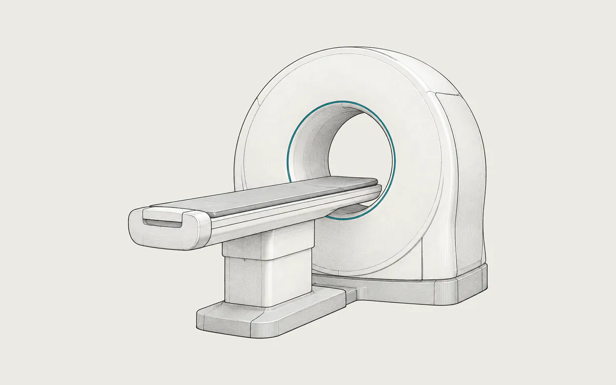
You lie on a table that moves through a ring-shaped opening called the gantry. Inside it, an X-ray tube and detectors rotate around the body several times per second. The detectors measure how much of the beam passes through the body from many directions, and a computer reconstructs images from these measurements. Modern scanners collect data while the table moves (helical scanning). From the same data they can reconstruct thin or thick slices and images in other planes.
Each pixel has a value in Hounsfield units (HU), which shows how strongly tissue absorbs X-rays compared with water. Water is 0 HU, air is about -1000 HU and fat is about -100 HU. Most soft tissues are between +20 and +80 HU, and bone ranges from a few hundred to more than +1000 HU.
Uses
Common uses include:
- injuries and internal bleeding after accidents
- stroke and other urgent brain problems
- lung diseases, such as pneumonia or blood clots in the lungs
- abdominal pain, such as appendicitis or kidney stones
- finding and staging cancer and checking the response to treatment
- fractures, blood vessels (CT angiography) and the heart
- guiding biopsies and planning radiation therapy
Preparation and what to expect
Before a scan with contrast, you may be asked not to eat for a few hours. Tell the staff if you are or might be pregnant, if you had a reaction to contrast before, and if you have kidney disease or diabetes. Remove metal objects from the area to be scanned.
You lie on the table, often with your arms above your head. The technologist works in the next room but can see and hear you. You may be asked to hold your breath for a few seconds. A contrast injection can cause a brief warm feeling or a metallic taste. The scan is painless. A radiologist then sends a report to your doctor.
Safety
CT uses ionizing radiation. RadiologyInfo lists typical effective doses for adults of about 1.6 mSv for the head, 6.1 mSv for the chest, 7.7 mSv for the abdomen and pelvis, and 1.5 mSv for low-dose lung cancer screening. Scanning with and without contrast can double the dose. Natural background radiation is about 3 mSv per year, so a chest CT equals about two years of background radiation. The actual dose depends on the protocol, the machine and the size of the patient.
Radiation at these levels may slightly increase the risk of cancer later in life. The risk from one scan is small. Children are more sensitive, so their scans use lower settings. You can ask the referring doctor why the scan is needed and whether a test without radiation, such as MRI or ultrasound, could answer the same question.
Serious allergic reactions to iodine-based contrast are rare. Contrast can affect the kidneys of people with reduced kidney function, so doctors may test kidney function first.
What a CT study looks like in DICOM
CT images have the value CT in Modality (0008,0060). Most scanners store each slice as a separate file of the SOP class CT Image Storage (1.2.840.10008.5.1.4.1.1.2). Some newer scanners use Enhanced CT Image Storage (1.2.840.10008.5.1.4.1.1.2.1), which can keep a whole series in one multi-frame file.
A typical slice has 512x512 pixels, with 16 bits allocated and 12 to 16 bits stored per pixel. The stored numbers are not Hounsfield units: a viewer multiplies each value by Rescale Slope (0028,1053) and adds Rescale Intercept (0028,1052). A slope of 1 and an intercept of -1024 are common, but vendors differ.
A study usually has several series, for example:
- localizer images (scout views) used to plan the scan
- axial slices reconstructed for soft tissue and, with a sharper kernel, for lungs or bone
- thin slices (0.5 to 1.25 mm) for computer processing and thicker slices (3 to 5 mm) for reading
- coronal and sagittal reformats
- contrast phases, such as arterial and venous
Useful tags include Slice Thickness (0018,0050), Convolution Kernel (0018,1210) with vendor-specific names, KVP (0018,0060), CTDIvol (0018,9345), Image Position (Patient) (0020,0032), Window Center (0028,1050) and Window Width (0028,1051).
Many scanners also save a dose report: an X-Ray Radiation Dose SR (1.2.840.10008.5.1.4.1.1.88.67) with the CTDIvol and dose-length product (DLP) of each acquisition, a Secondary Capture image (1.2.840.10008.5.1.4.1.1.7) showing the dose table, or both.
How to view the images
In a DICOM viewer you can scroll through a series, measure values in HU and change the window, the range of values shown as shades of gray. Typical presets are lung (width 1500, center -600), soft tissue (width 400, center 40) and bone (width 1800, center 400). Hospitals use slightly different values.
Multiplanar reconstruction (MPR) shows the slices in other planes and works best with thin slices. Weasis (free, open source), Horos (free, open source, macOS) and RadiAnt (commercial, Windows) support MPR, and 3D Slicer (free, open source) adds advanced 3D tools. See how to open DICOM files, and remove personal data before you share images publicly.