ModalityIOPXCT
Dental X-rays and CBCT
Small intraoral images, one panoramic view of both jaws, and 3D cone beam CT. Many dental systems use their own formats and export DICOM on request.
On this page
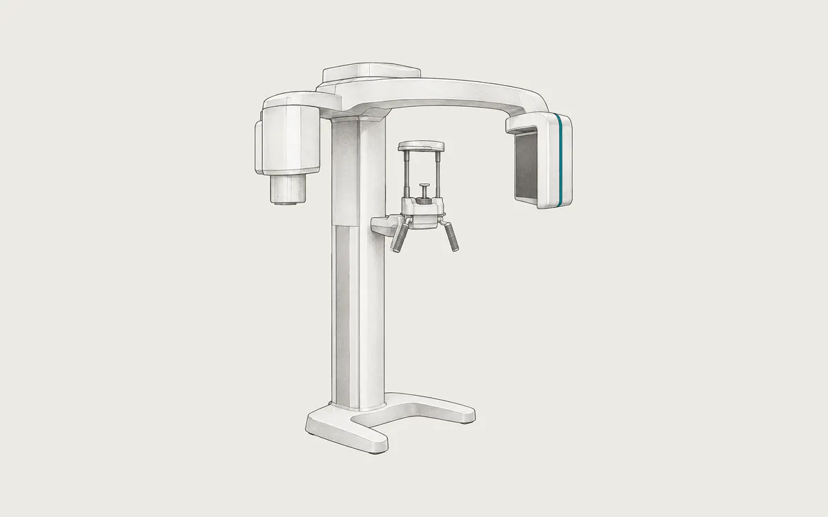
Dentists use several kinds of X-ray images. Intraoral images are taken with a small sensor inside the mouth and show a few teeth in fine detail. A panoramic image shows all teeth and both jaws in one wide picture. Cone beam computed tomography (CBCT) makes a 3D image of the teeth, jaws and face.
In DICOM, these images have three modality codes: IO for intraoral, PX for panoramic and CT for CBCT. Many dental programs store images in their own formats and create DICOM files only on export.
Key facts
| Fact | Details |
|---|---|
| DICOM modality code | IO (intraoral), PX (panoramic), CT (CBCT) |
| Typical number of images | 2 to 4 bitewings, 18 to 20 for a full-mouth series, 1 panoramic, hundreds of CBCT slices |
| Typical study size | 1 to 10 MB per 2D image; about 50 to 500 MB for CBCT |
| Exam duration | Seconds per intraoral image; up to about 40 seconds for a panoramic or CBCT scan |
| Radiation | Yes, low; about 0.005 mSv intraoral, 0.025 mSv panoramic, 0.18 mSv CBCT |
| Contrast agent | None |
How it works
Intraoral images
A small digital sensor or a phosphor plate is placed in the mouth, and the X-ray tube is aimed from outside the cheek. Bitewing images show the crowns of upper and lower back teeth and help to find decay between teeth. Periapical images show whole teeth, including the roots and the surrounding bone.
Panoramic images
The X-ray tube and the detector move around the head for about 10 to 20 seconds. The result is one flat image of both jaws, all teeth, the jaw joints and part of the sinuses. Some areas are slightly blurred or enlarged, so the image is less suitable for exact measurements.
Cone beam CT
A cone-shaped X-ray beam and a flat detector rotate once around the head and record many 2D images from different angles. A computer reconstructs a 3D volume made of small cubes called voxels, often 0.075 to 0.4 mm in size. The field of view can cover a few teeth or the whole face. CBCT shows teeth and bone well but soft tissues poorly. Unlike medical CT, its gray values are not reliable Hounsfield units.
Uses
- tooth decay, especially between teeth
- bone loss from gum disease
- infections at the root tips and root canal treatment
- impacted teeth, such as wisdom teeth
- orthodontic planning
- planning dental implants and jaw surgery, often with CBCT
- injuries, cysts and tumors of the jaws
Dentists decide how often X-rays are needed based on each person's risk, not on a fixed schedule.
Preparation and what to expect
No special preparation is needed. Remove glasses, earrings, piercings, hairpins and removable dentures, because metal disturbs the images. Tell the dentist if you are or might be pregnant.
For intraoral images you hold the sensor in place by biting on a holder. For panoramic and CBCT scans you stand or sit, bite on a small block and rest your chin on a support. Stay completely still while the machine moves around your head, because movement blurs the images. The scan is painless.
Safety
Dental X-rays use low doses. RadiologyInfo lists about 0.005 mSv for a dental X-ray (about one day of natural background radiation), 0.025 mSv for a panoramic X-ray (about three days) and 0.18 mSv for a cone beam CT (about 22 days). Natural background radiation is about 3 mSv per year. CBCT doses vary widely with the field of view and the settings, so CBCT is used only when 2D images are not enough. The dose depends on the protocol, the machine and body size. Children are more sensitive to radiation, so their settings should be lower.
The American Dental Association no longer recommends thyroid collars for dental imaging, because they can block the beam and cause repeat images. Rules in other countries may differ. Needed dental X-rays are generally considered safe during pregnancy, but always tell the dentist.
What dental images look like in DICOM
- Intraoral images use Digital Intra-Oral X-Ray Image Storage - For Presentation (
1.2.840.10008.5.1.4.1.1.1.3) with ModalityIO, one file per image. Images are often 1 to 4 megapixels with 8 to 16 bits per pixel. The file can record which teeth are shown, as coded entries in Primary Anatomic Structure Sequence(0008,2228), along with Anatomic Region Sequence(0008,2218)and Image Laterality(0020,0062). - Panoramic images usually use Digital X-Ray Image Storage - For Presentation (
1.2.840.10008.5.1.4.1.1.1.1) with ModalityPX. Older and plate-based systems may use Computed Radiography Image Storage (1.2.840.10008.5.1.4.1.1.1). A panoramic image is often about 2500 to 3000 pixels wide. - CBCT volumes usually use CT Image Storage (
1.2.840.10008.5.1.4.1.1.2) with ModalityCT, one file per slice: a few hundred slices of 400x400 to 800x800 pixels with 16 bits allocated. Some programs export one multi-frame file instead, such as Enhanced CT Image Storage (1.2.840.10008.5.1.4.1.1.2.1). Pixel Spacing(0028,0030)and Slice Thickness(0018,0050)give the voxel size.
DICOM export is often an option that must be chosen, and some clinics give out JPEG files by default. A JPEG copy loses bit depth and most metadata, including the pixel size needed for measurements. For treatment in another clinic, ask for a DICOM export. CBCT discs often include the vendor's own viewer.
How to view the images
Intraoral and panoramic images open in any DICOM viewer. CBCT needs a viewer with multiplanar reconstruction (MPR) to show the volume in three planes. Weasis (free, open source) can also build a panoramic view from a CBCT volume along a curve drawn on the dental arch. Horos (free, open source, macOS), 3D Slicer (free, open source) and RadiAnt (commercial, Windows) show CBCT in several planes and in 3D. Implant planning uses dedicated dental software. See how to open DICOM files.