ModalityBMD
Bone density scan (DXA)
A low-dose X-ray scan that measures bone mineral density in the spine and hip. DICOM exports are often report images rather than raw scan data.
On this page
A bone density scan measures how much mineral, mainly calcium, is in your bones. The standard method is dual-energy X-ray absorptiometry, called DXA or DEXA. It is usually done on the lower spine and the hips and is used to diagnose osteoporosis, a disease in which bones become thin and break easily.
In DICOM, bone densitometry has the modality code BMD. A DXA study is small: usually a few report images, sometimes with a structured report that holds the numbers.
Key facts
| Fact | Details |
|---|---|
| DICOM modality code | BMD |
| Typical number of images | 1 to 10 objects, mostly report pages |
| Typical study size | 1 to 20 MB |
| Exam duration | 10 to 30 minutes |
| Radiation | Yes, very low; about 0.001 mSv |
| Contrast agent | None |
How it works
The DXA machine sends a thin beam of low-dose X-rays with two different energies through the body. Bone and soft tissue absorb the two energies differently. By comparing the two measurements, the machine removes the effect of soft tissue and calculates the amount of bone mineral. It divides this amount by the bone area and reports bone mineral density (BMD) in grams per square centimeter (g/cm²).
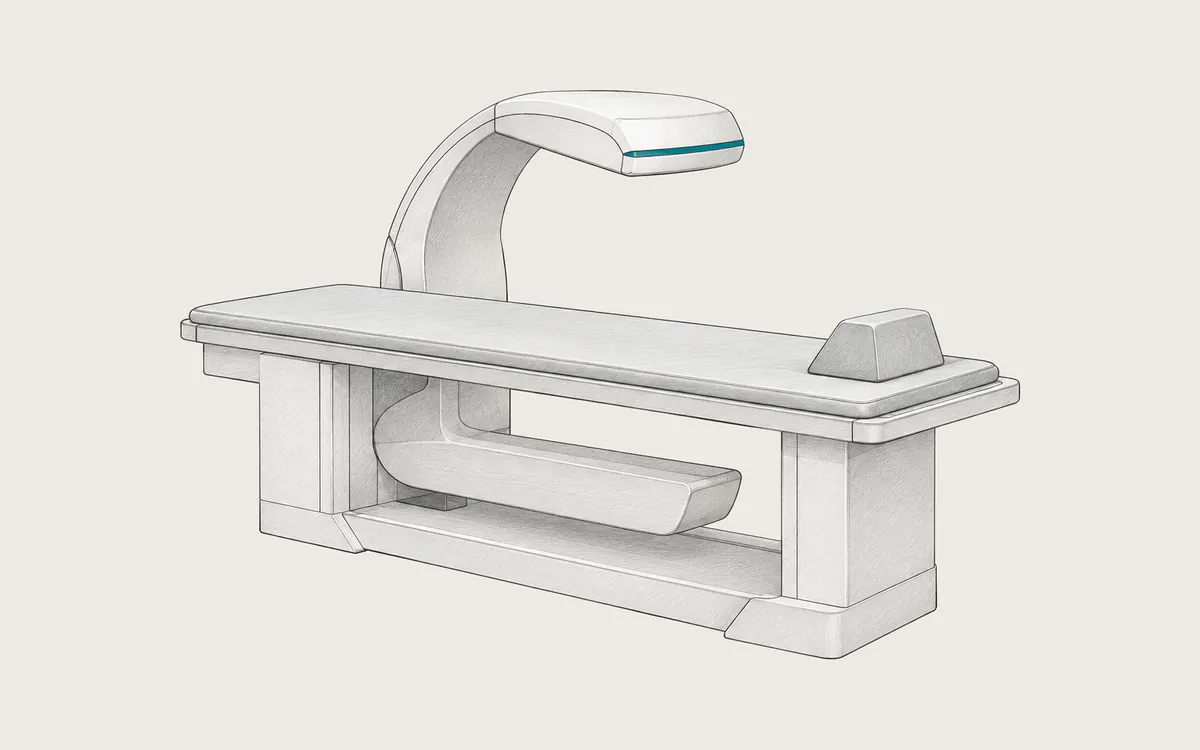
The X-ray source is under a padded table, and a detector in an arm above it moves slowly over the area being measured. The usual regions are the lumbar spine, the total hip and the femoral neck; sometimes the forearm is added. Whole-body scans can also measure body composition: fat, lean tissue and bone. Small devices for the wrist or heel are mainly used for screening.
Many machines can also take a side image of the spine to find vertebral fractures (vertebral fracture assessment, VFA).
Uses
- diagnosing osteoporosis and low bone mass
- estimating the risk of fractures, often together with risk calculators such as FRAX
- monitoring treatment, usually with scans one to two years apart
- checking people who take bone-weakening medicines, such as long-term corticosteroids, or have diseases that cause bone loss
T-score and Z-score
The report gives the BMD of each region and two scores.
The T-score compares your BMD with the average of healthy young adults of the same sex. It is used for women after menopause and men aged 50 and older. The World Health Organization classification is:
- normal: -1.0 or higher
- low bone mass, also called osteopenia: between -1.0 and -2.5
- osteoporosis: -2.5 or lower
The Z-score compares your BMD with people of your own age and sex. It is used for children, women before menopause and men under 50. A Z-score of -2.0 or lower is described as below the expected range for age.
Bone density is only one part of fracture risk, so your doctor interprets it together with age, earlier fractures and other factors.
Preparation and what to expect
You can eat normally on the day of the exam, but do not take calcium supplements for at least 24 hours before it. Wear loose clothes without metal zippers, buttons or belts. If you recently had a barium study, a CT scan with contrast or a nuclear medicine scan, tell the staff: leftover material can distort the result, and you may need to wait 10 to 14 days. Tell them if you are or might be pregnant.
You lie on your back on the padded table. Your legs rest on a padded block for the spine, and your foot is held in a brace for the hip. You must lie still and may need to hold your breath for a few seconds. The scan is painless.
Safety
The radiation dose of DXA is very low. RadiologyInfo lists about 0.001 mSv, similar to about three hours of natural background radiation (about 3 mSv per year) and less than one-tenth of the dose of a chest X-ray. The exact value depends on the machine, the regions scanned and body size, but it stays very small. A VFA image or a whole-body scan adds a small extra dose. No contrast agent or tracer is used, and no radiation stays in the body.
DXA is not recommended during pregnancy. It does not affect breastfeeding.
What a DXA study looks like in DICOM
DXA systems differ widely in what they send as DICOM. The full scan data used for analysis usually stays in the vendor's own database on the DXA workstation. Common exports are:
- report pages saved as secondary capture images (
1.2.840.10008.5.1.4.1.1.7), with the scan images, BMD values, T-scores, Z-scores and trend graphs drawn into the pixels. Their Modality is usuallyBMD, but some systems use other values, such asOT. - a structured report with the measured values as data that a reporting system can read. Its Modality is always
SR, and the codes inside are often vendor-specific. - sometimes an encapsulated PDF report (
1.2.840.10008.5.1.4.1.1.104.1).
Numbers drawn into report images are part of the picture, so they cannot be searched or recalculated. The reference data for T-scores and Z-scores depend on sex, age and sometimes ethnicity, so Patient's Sex (0010,0040) and Patient's Birth Date (0010,0030) must be correct.
Results from different machines cannot be compared directly, so follow-up scans should be done at the same place, ideally on the same machine.
How to view the images
Secondary capture report pages open in almost any DICOM viewer, for example Weasis (free, open source), MicroDicom (free for non-commercial use, Windows) or RadiAnt (commercial, Windows). Structured reports are shown less consistently: some viewers show the values, others ignore them, so the PDF or printed report may be easier to read. A general viewer cannot redo the bone density analysis, which needs the vendor's DXA software. See how to open DICOM files.