ModalityXARF
Angiography and fluoroscopy
Live X-ray imaging of blood vessels, the heart and the digestive tract. Each recorded run is one multi-frame DICOM file that plays like a short video.
On this page
Fluoroscopy is real-time X-ray imaging that shows moving images on a monitor, like an X-ray video. Angiography uses it with a contrast agent to show blood vessels. In catheter angiography, a doctor guides a thin tube (catheter) through a blood vessel and injects contrast through it. The same equipment is used for treatment, for example to open a narrowed artery with a stent.
In DICOM, angiography images have the modality code XA and fluoroscopy images the code RF. Each recorded sequence, called a run, is usually one multi-frame file.
Key facts
| Fact | Details |
|---|---|
| DICOM modality code | XA for angiography; RF for fluoroscopy |
| Typical number of images | 5 to 50 runs, each with 10 to several hundred frames |
| Typical study size | 100 MB to 2 GB |
| Exam duration | About 20 minutes for a barium study; up to several hours for angiography |
| Radiation | Yes; varies widely: often several mSv, more for long procedures |
| Contrast agent | Iodine-based contrast; barium or iodine-based contrast for digestive studies |
How it works
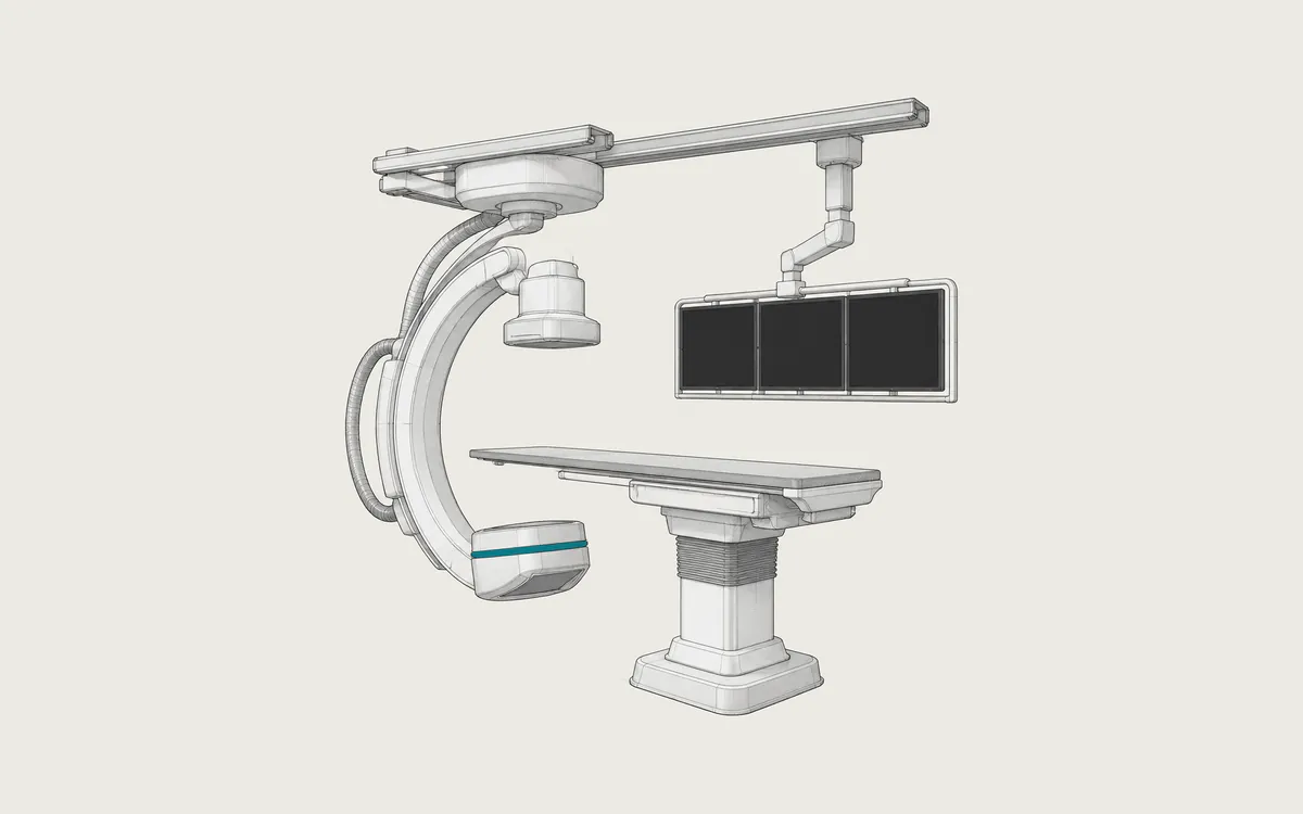
The X-ray tube and the detector sit on opposite ends of a C-shaped arm (C-arm) that rotates around the patient. Most systems use pulsed fluoroscopy, short bursts of X-rays, to reduce the dose. The doctor watches live images while moving the catheter. Only some sequences are recorded as runs, with a higher dose per frame and clearer images; most live fluoroscopy is not saved.
Outside the heart, digital subtraction angiography (DSA) is common. The system takes a mask image before the contrast arrives and subtracts it from later frames, so bones and other tissues disappear and only the vessels filled with contrast remain. The heart moves all the time, so heart runs are usually recorded without subtraction. A rotational run can be reconstructed into a 3D image of the vessels.
Uses
- coronary angiography and stents for chest pain or a heart attack
- brain angiography for aneurysms and removing clots in stroke
- arteries of the legs, kidneys and other organs
- embolization: blocking a bleeding vessel or the blood supply of a tumor
- barium studies of swallowing, the stomach and the bowel
- pacemakers, joint injections and guidance during some surgeries
Preparation and what to expect
If you will receive a sedative, you may need to stop eating and drinking four to eight hours before. A blood test checks kidney function and blood clotting. Tell the staff about your medicines, especially blood thinners and diabetes medicines, and about any allergy to iodine-based contrast. After sedation, someone must drive you home.
The doctor numbs the skin at the groin or the wrist and inserts the catheter through a small cut. You do not feel the catheter inside the vessels. When contrast is injected, you may feel warm for a few seconds. Afterwards the doctor removes the catheter and presses on the site, or uses a closure device. You may need to lie flat for several hours.
Barium studies are simpler: you drink the contrast liquid and change position on the table while the radiologist takes images.
Safety
Radiation doses depend strongly on how long the procedure takes. In a European study at several centers, the median effective dose of a diagnostic coronary angiography was 7.4 mSv, with large differences between centers. This is about two and a half years of natural background radiation (about 3 mSv per year). Complex treatments give higher doses. Very long procedures can rarely injure the skin where the beam enters the body. The dose depends on the protocol, the machine, the procedure time and body size.
Serious allergic reactions to iodine-based contrast are rare. Contrast can harm the kidneys, mainly in people with kidney disease or diabetes. The catheter can cause bleeding or bruising and, rarely, damage to the artery or a blood clot.
Tell the staff if you are or might be pregnant. If you are breastfeeding, ask your doctor; the American College of Radiology reports that very little contrast reaches the baby.
What an angiography study looks like in DICOM
Angiography runs use X-Ray Angiographic Image Storage (1.2.840.10008.5.1.4.1.1.12.1) with Modality XA. Fluoroscopy uses X-Ray Radiofluoroscopic Image Storage (1.2.840.10008.5.1.4.1.1.12.2) with Modality RF, but the two do not always match: a fluoroscopy system may store XA objects with Modality RF. Newer systems may use Enhanced XA Image Storage (1.2.840.10008.5.1.4.1.1.12.1.1), and 3D reconstructions may use X-Ray 3D Angiographic Image Storage (1.2.840.10008.5.1.4.1.1.13.1.1).
Number of Frames (0028,0008) gives the frame count, and Frame Time (0018,1063) the time between frames in milliseconds; runs with a changing rate use Frame Time Vector (0018,1065). Heart runs are usually recorded at 7.5 to 30 frames per second, subtraction runs often at lower rates. Frames are usually 512x512 or 1024x1024 pixels with 8 to 12 bits stored. Lossless JPEG compression is common.
Other useful tags:
- Positioner Primary Angle
(0018,1510): from right anterior oblique (RAO, negative) to left anterior oblique (LAO, positive) - Positioner Secondary Angle
(0018,1511): toward the feet (caudal, negative) or the head (cranial, positive) - Mask Subtraction Sequence
(0028,6100): the DSA mask frames; some systems save subtracted runs separately - Image and Fluoroscopy Area Dose Product
(0018,115E): the dose area product of the run and the fluoroscopy before it
Many systems also save an X-Ray Radiation Dose SR (1.2.840.10008.5.1.4.1.1.88.67) with the total fluoroscopy time, dose area product and air kerma of the whole procedure, including fluoroscopy that was not saved as images.
How to view the images
A viewer must play each run as a loop at the recorded frame rate. Weasis (free, open source), Horos (free, open source, macOS), MicroDicom (free for non-commercial use, Windows) and RadiAnt (commercial, Windows) can play XA runs. The image is magnified, so vessel sizes measured in a general viewer may not be exact. See how to open DICOM files.