ModalityNM
Nuclear medicine scans and SPECT
A gamma camera records a radioactive tracer to show how organs work. A study has only a few DICOM files, but one file can hold hundreds of frames.
On this page
Nuclear medicine scans, also called scintigraphy, show how organs work. A small amount of a radioactive tracer is injected, swallowed or inhaled. It collects in an organ or tissue, and a gamma camera records the gamma rays it gives off. SPECT (single photon emission computed tomography) is the 3D form of this test, and SPECT/CT adds a CT scan.
In DICOM, all of these use the modality code NM, and one file can hold many images, called frames.
Key facts
| Fact | Details |
|---|---|
| DICOM modality code | NM (also SPECT); CT for the CT part of SPECT/CT |
| Typical number of images | 1 to 20 files, each with 1 to several hundred frames |
| Typical study size | 1 to 50 MB; more with a CT |
| Exam duration | 20 minutes to several hours; some tests take several days |
| Radiation | Yes, from the tracer; about 1 to 6 mSv for most routine scans |
| Contrast agent or tracer | A radioactive tracer, most often technetium-99m |
How it works
Most scans use technetium-99m, which has a half-life of about 6 hours. It is attached to carrier molecules that go to specific targets, such as bone, heart muscle or kidneys.
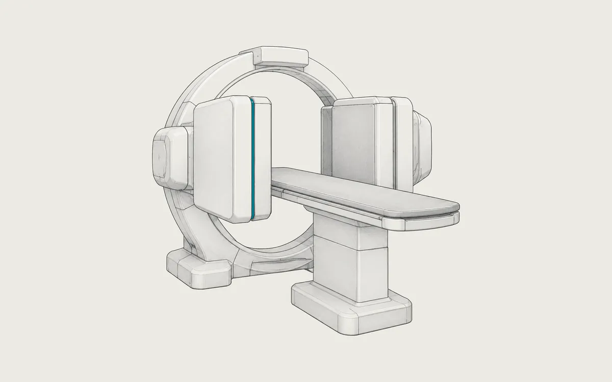
A gamma camera has a large detector behind a lead collimator, a plate with small holes that passes only gamma rays traveling straight toward it. Cameras record static views, dynamic series over time, heart images synchronized with the electrocardiogram (ECG) and whole-body images.
For SPECT, the detectors rotate around the patient and record many views from different angles, often 60 to 128. A computer reconstructs slices from these views. In SPECT/CT, the CT shows anatomy and corrects for gamma rays absorbed in the body.
Uses
- bone scans for fractures, infection and cancer that has spread to bone
- blood flow to the heart muscle, at rest and during a stress test
- thyroid, parathyroid, lung and kidney scans
- gallbladder function and bleeding in the bowel
- brain scans, for example for Parkinson's disease
- finding the sentinel lymph node before cancer surgery
Nuclear medicine also treats some diseases; images taken after treatment are stored the same way.
Preparation and what to expect
Preparation depends on the test. Before a heart stress test you may need to avoid caffeine and smoking for up to 48 hours and not eat for several hours. Before thyroid tests you may need to stop some medicines and avoid iodine. For a bone scan you usually drink water and empty your bladder often. Tell the staff about all your medicines.
After the tracer is given, there is often a wait: minutes for some tests, 2 to 4 hours for a bone scan, and up to several days for some tracers. During imaging the detector heads come very close to your body, but the camera itself gives off no radiation. You must keep still, often for 20 minutes or longer. Young children may need gentle wrapping or sedation. Usually you can return to normal activities right away.
Safety
The radiation comes from the tracer inside your body. The UK Health Security Agency lists typical effective doses of about 1 mSv for lung, kidney and thyroid scans, 4 mSv for a bone scan and 6 mSv for a technetium-99m heart scan. An older heart test with thallium-201 gives about 18 mSv. Natural background radiation is about 3 mSv per year, depending on where you live. The CT in SPECT/CT adds its own dose. The dose depends on the test, the tracer activity, the machine and body size.
Allergic reactions to tracers are extremely rare. With technetium-99m, most of the radioactivity is gone within a day; drinking water and emptying your bladder often helps. After treatments, you receive rules about contact with other people.
During pregnancy, a scan is done only when the benefit outweighs the risk, so tell the staff if you might be pregnant. If you are breastfeeding, tell them before the test, because some tracers pass into breast milk and a pause may be needed.
What a nuclear medicine study looks like in DICOM
Gamma camera and SPECT images have the value NM in Modality (0008,0060); the old SPECT code ST is retired. They use Nuclear Medicine Image Storage (1.2.840.10008.5.1.4.1.1.20), a multi-frame object. One file holds a whole acquisition, such as all time frames of a kidney study or all projections of a SPECT scan. All frames in one file have the same matrix size.
The third value of Image Type (0008,0008) tells what the file contains: STATIC, DYNAMIC, GATED, WHOLE BODY, TOMO (SPECT projections), GATED TOMO, RECON TOMO (reconstructed slices) or RECON GATED TOMO. Number of Frames (0028,0008) gives the frame count. Frame Increment Pointer (0028,0009) names the vectors that describe each frame, such as Detector Vector (0054,0020), Angular View Vector (0054,0090) or Slice Vector (0054,0080).
Matrices are small: 64x64 or 128x128 for SPECT projections, 128x128 or 256x256 for static images and often 256x1024 for whole-body images, with 8 or 16 bits per pixel. In NM files, Radionuclide Total Dose (0018,1074) in the Radiopharmaceutical Information Sequence (0054,0016) is in megabecquerels (MBq), while PET files use becquerels.
Workstations often save results, such as heart function charts or kidney curves, as secondary capture images (1.2.840.10008.5.1.4.1.1.7). These are pictures of a screen and cannot be reprocessed. In SPECT/CT, the CT images form a separate CT series in the same study.
How to view the images
A viewer must show the frames of a multi-frame file in the right order, as a cine loop, a set of views or a stack of slices. Not all general viewers do this well. Weasis (free, open source), Horos (free, open source, macOS) and RadiAnt (commercial, Windows) can open NM images, and Weasis can overlay SPECT on CT. Heart analysis needs dedicated software. See how to open DICOM files.