Imaging studies stored in DICOM
Ten common medical imaging studies: how each one is done, what it shows and what its DICOM files contain.
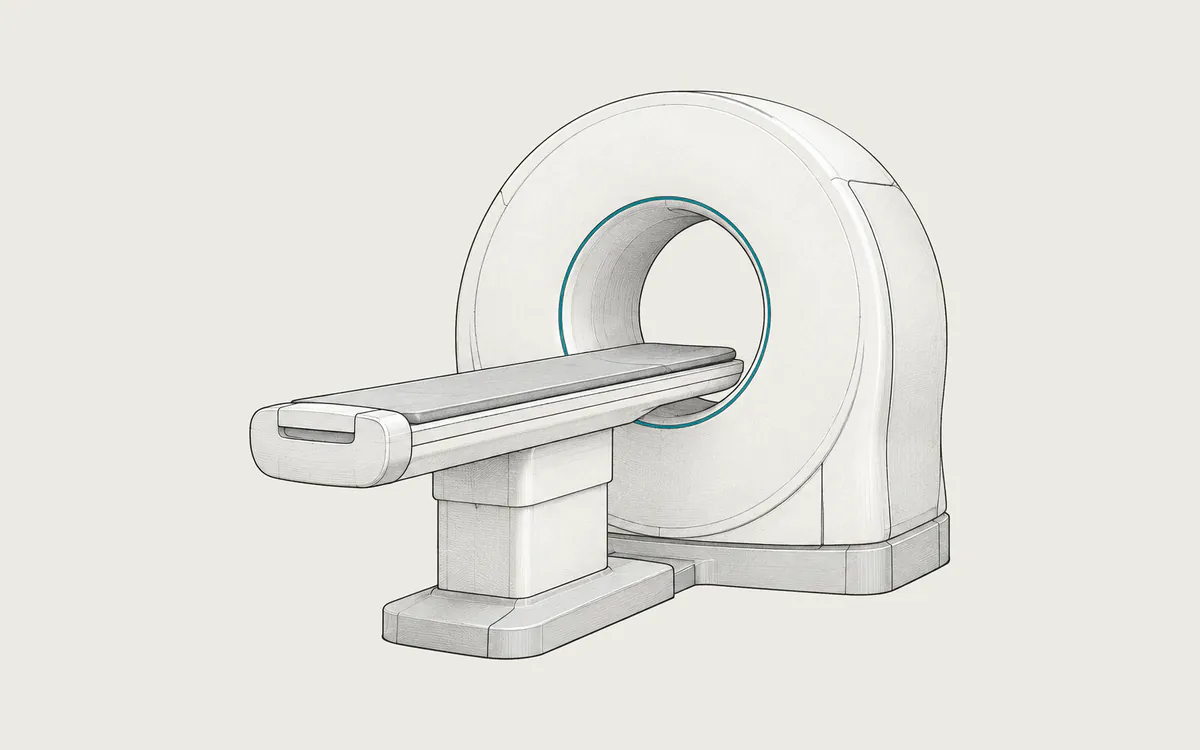
CT
CT
Cross-sectional X-ray imaging of any part of the body. One study holds hundreds to thousands of DICOM slices with values in Hounsfield units.
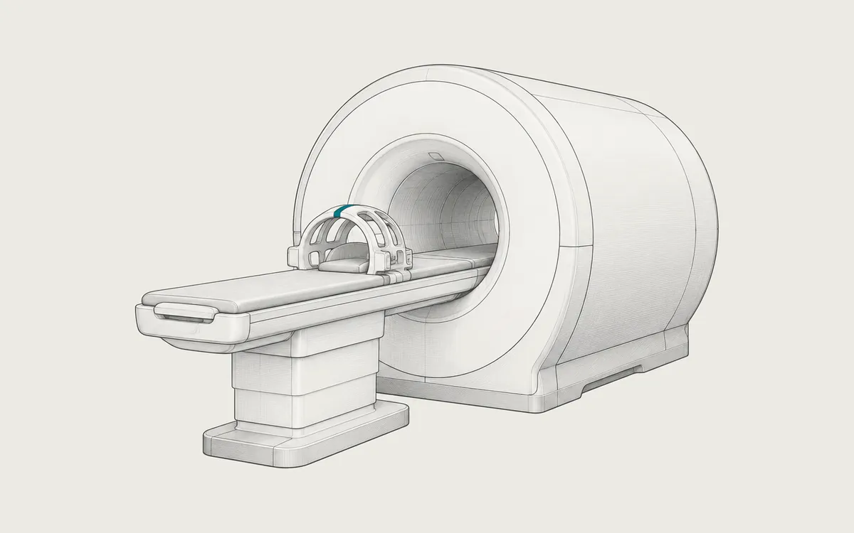
MR
MRI
Imaging with a strong magnet and radio waves, without X-rays. Each sequence is a separate DICOM series, so one study often has hundreds or thousands of images.
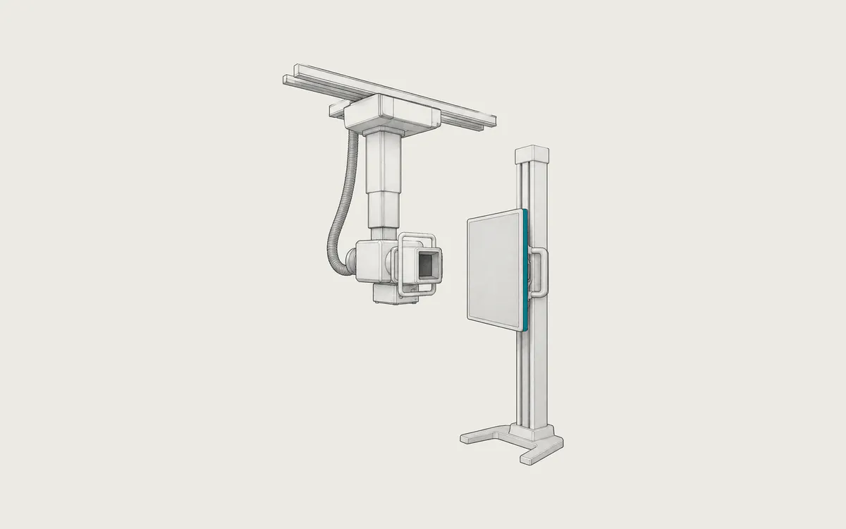
CRDX
X-ray
A projection image made with a very short X-ray exposure. A study usually has one to four large grayscale DICOM images.
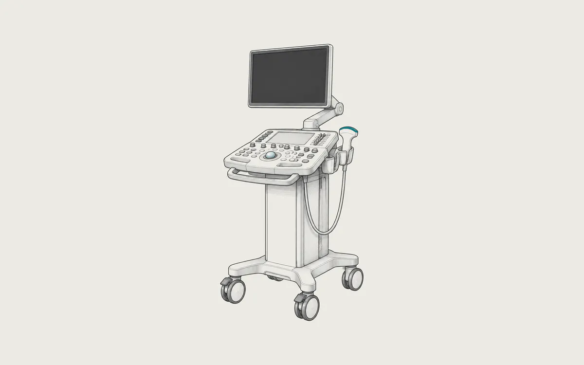
US
Ultrasound
Real-time imaging with high-frequency sound waves and no radiation. A study mixes still images with video clips (cine loops), which are stored as multi-frame DICOM files.
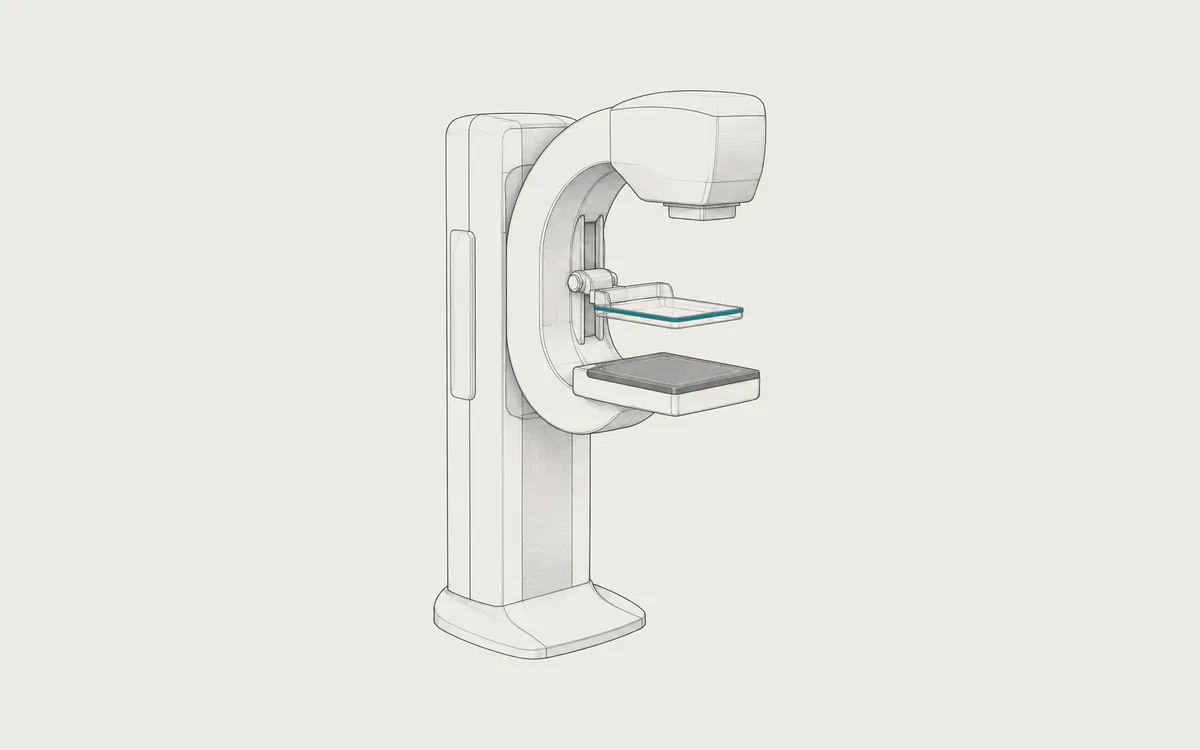
MG
Mammography
Low-dose X-ray imaging of the breast. A standard study has four very large images; tomosynthesis adds stacks of thin slices and can reach several gigabytes.
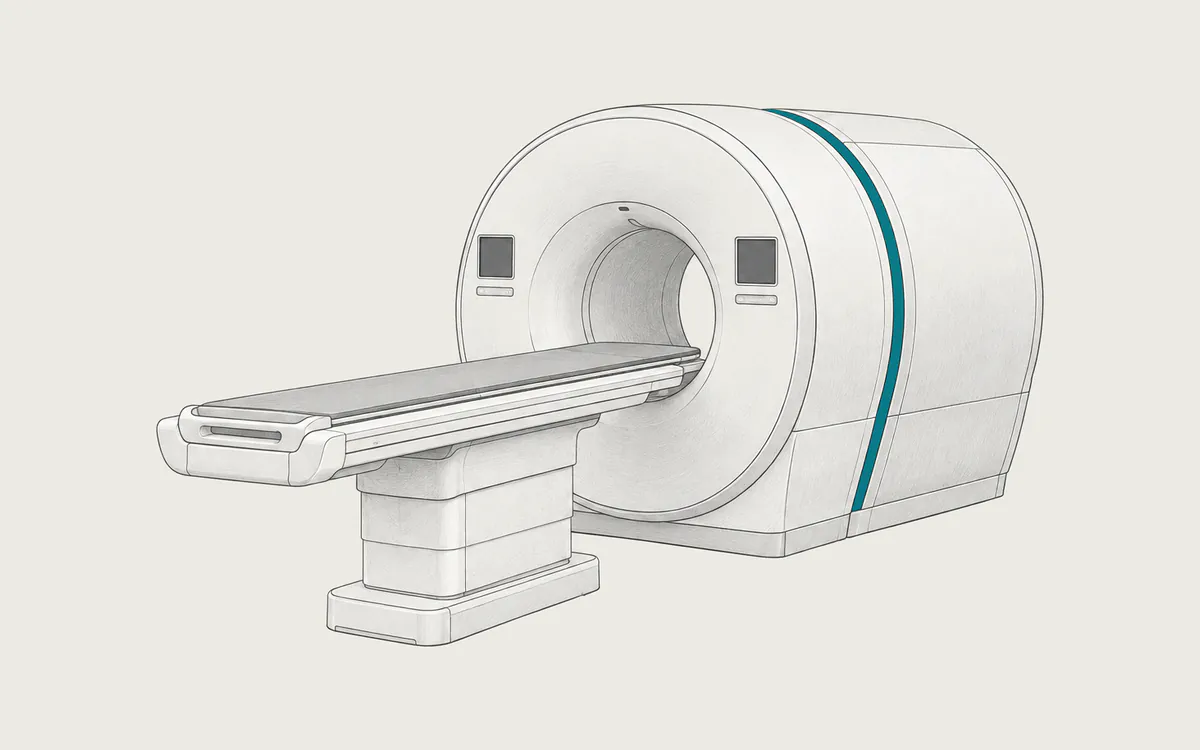
PT
PET/CT
A radioactive tracer shows how active tissues are, and CT shows where they are. A viewer can fuse the PET (PT) and CT series into one picture.
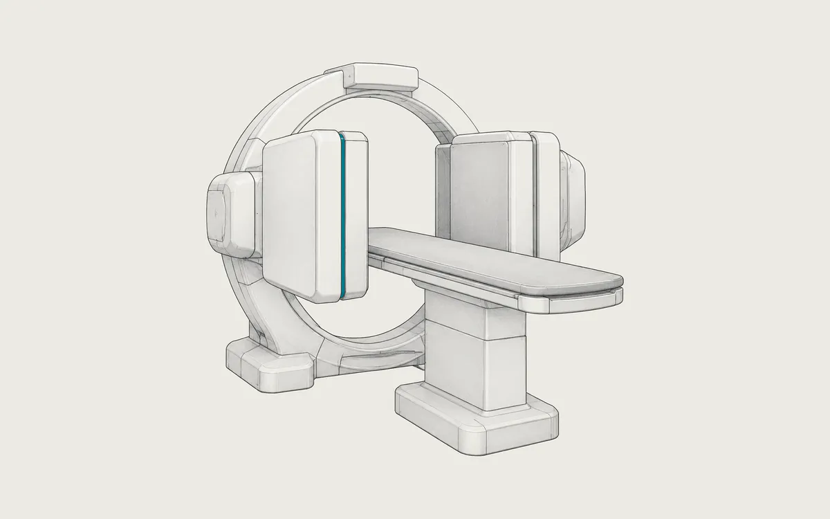
NM
Nuclear medicine
A gamma camera records a radioactive tracer to show how organs work. A study has only a few DICOM files, but one file can hold hundreds of frames.
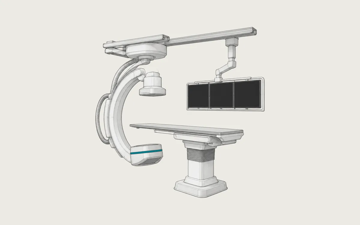
XARF
Angiography
Live X-ray imaging of blood vessels, the heart and the digestive tract. Each recorded run is one multi-frame DICOM file that plays like a short video.
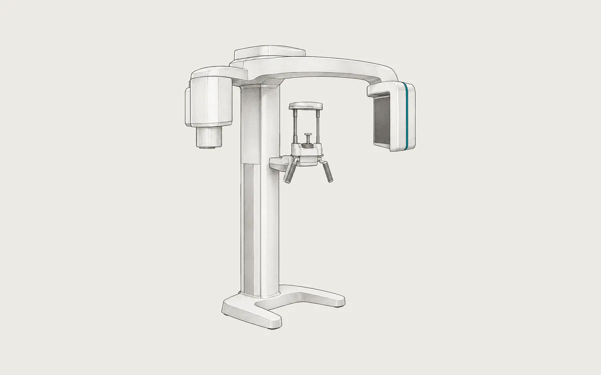
IOPXCT
Dental
Small intraoral images, one panoramic view of both jaws, and 3D cone beam CT. Many dental systems use their own formats and export DICOM on request.
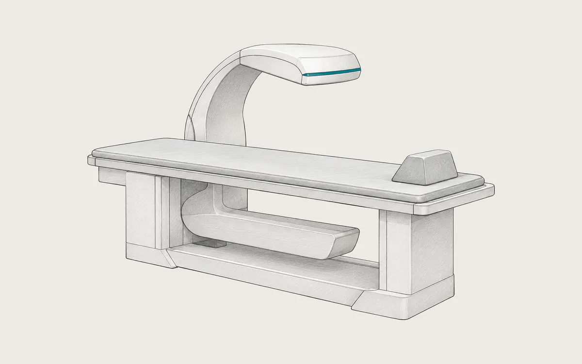
BMD
DXA
A low-dose X-ray scan that measures bone mineral density in the spine and hip. DICOM exports are often report images rather than raw scan data.