ModalityMR
MRI (magnetic resonance imaging)
Imaging with a strong magnet and radio waves, without X-rays. Each sequence is a separate DICOM series, so one study often has hundreds or thousands of images.
On this page
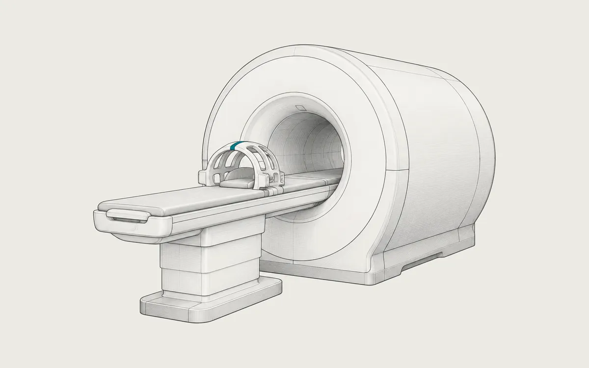
MRI (magnetic resonance imaging) uses a strong magnet, radio waves and a computer to make detailed images of the inside of the body. Unlike CT, it does not use X-rays. MRI shows soft tissues especially well, such as the brain, the spinal cord, joints and many organs.
Key facts
| Fact | Details |
|---|---|
| DICOM modality code | MR |
| Typical number of images | Several hundred to a few thousand, in 5 to 15 or more series |
| Typical study size | 50 MB to 1 GB or more |
| Exam duration | Usually 15 to 60 minutes, sometimes longer |
| Radiation | None (no ionizing radiation) |
| Contrast agent | Gadolinium-based, injected into a vein, in some exams |
How it works
The scanner is a large tube with a very strong magnet, usually 1.5 or 3 tesla. Hydrogen nuclei (protons) in the water and fat of the body line up with the magnetic field. Short radio pulses tip the protons out of alignment. As they return, the protons send out weak radio signals, which coils near the body receive. Gradient coils vary the magnetic field slightly so that the computer can locate each signal. Their rapid switching causes the loud knocking noise.
Different timings of the pulses produce different sequences, such as T1-weighted, T2-weighted, FLAIR and diffusion-weighted images. A tissue can look bright in one sequence and dark in another, and this helps doctors tell tissues apart. Compared with CT, an MRI exam takes longer and is more sensitive to movement.
Uses
Common uses include:
- the brain, spinal cord and spine, for example in stroke, multiple sclerosis, tumors or disc problems
- joints, ligaments, cartilage and muscles after injuries
- the liver, pancreas, kidneys, prostate, uterus and other organs
- the heart and blood vessels (MR angiography)
- the breasts of some patients, in addition to mammography
Preparation and what to expect
Before the scan, you fill in a safety questionnaire. Tell the staff about any implant or device, such as a pacemaker, defibrillator, cochlear implant, medication pump or aneurysm clip, and about metal fragments in your body, especially near the eyes. Bring your implant card if you have one. Also tell the staff if you are pregnant, have kidney disease or feel anxious in closed spaces.
You remove all metal objects, such as jewelry, watches, hearing aids, phones and bank cards. You often change into a gown, because some clothing contains metal threads. You lie on a table, a coil is placed around the body part, and the table moves into the tube. The scanner is loud, so you get earplugs or headphones. You need to lie still and sometimes hold your breath. You can talk to the technologist and use a call button.
Safety
MRI uses no ionizing radiation. The main risks come from the magnet, which is always on, and from the radio waves:
- Objects made of iron or steel can be pulled into the scanner with great force.
- Some implants can move, heat up or stop working. Devices are labeled MR Safe, MR Conditional (safe only under specific conditions) or MR Unsafe. Many modern pacemakers are MR Conditional. A device with unknown status is treated as MR Unsafe.
- Radio waves warm the body slightly. Wires, metal in clothing and some tattoos can rarely cause burns.
- The noise can harm hearing without ear protection.
Gadolinium-based contrast agents rarely cause allergic reactions. In people with severe kidney disease, some older agents were linked to a rare but serious condition called nephrogenic systemic fibrosis (NSF), so kidney function may be checked first. Small amounts of gadolinium can remain in the brain and other tissues for months or years. Harmful effects of this have not been confirmed in people with normal kidney function. Most centers avoid contrast during pregnancy. If you have concerns, discuss them with the referring doctor.
What an MRI study looks like in DICOM
MRI images have the value MR in Modality (0008,0060). Many scanners store each image as a separate file of the SOP class MR Image Storage (1.2.840.10008.5.1.4.1.1.4). Newer scanners can use Enhanced MR Image Storage (1.2.840.10008.5.1.4.1.1.4.1), which can keep a whole series in one multi-frame file. Some scanners use this form by default.
Typical matrices are 256x256 to 512x512 pixels, often interpolated from a smaller acquired matrix. Pixels have 16 bits allocated, and often 12 bits are used. Unlike CT, MRI pixel values have no absolute unit. Brightness depends on the sequence, the coil and the scanner, so values cannot be compared directly between scans. Calculated maps, such as ADC maps, are an exception.
A sequence can be repeated in several planes, each as its own series. A brain study may contain a localizer, sagittal T1, axial T2, FLAIR, diffusion with an ADC map, and T1 after contrast. Series Description (0008,103E) holds names such as AX T2 FLAIR, which differ between vendors and hospitals.
Useful tags include Repetition Time (0018,0080) and Echo Time (0018,0081) in milliseconds, Inversion Time (0018,0082), Magnetic Field Strength (0018,0087) in tesla, Slice Thickness (0018,0050) and Image Orientation (Patient) (0020,0037). Orientation matters because MRI slices are often oblique. Enhanced MR files store the diffusion b-value in a standard tag, Diffusion b-value (0018,9087); classic files often keep it in private tags.
How to view the images
In a DICOM viewer, you can open several series side by side and link their scrolling. MRI has no fixed window presets: the viewer starts with the window stored in the file, and you can adjust it.
Weasis (free, open source), Horos (free, open source, macOS) and OHIF Viewer (free, open source, web browser) can display MRI. If a viewer cannot open a series or shows only one image of it, the series may be an Enhanced MR file that the viewer does not support. See how to open DICOM files.